![An Australian Government Initiative [logo]](/images/austgovt_brown_90px.gif)

Case StudiesApical CellsGrowth of bryophyte gametophytes is under control of apical cells. An apical cell is a cell that divides repeatedly to form new cells. An apical cell has a front face and up to three rear faces. All cell division takes place along the rear faces and never along the front face. The concept of an apical cell is probably best explained by means of a very simple and artificial two-dimensional example as shown in the following sequence of diagrams.
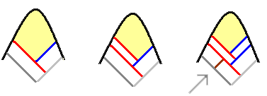
The leftmost diagram shows an apical cell, in colour, at the tip of a stem of a leafy bryophyte. The front face is curved and there are two, straight rear faces. These three faces delimit the apical cell. Behind the rear faces there are of course the cells of the rest of the stem, but they are irrelevant. In the next diagram on the first row the red line indicates a new cell wall that has been formed, parallel to the left rear face of the apical cell. That means one new cell has been formed from part of the original apical cell. The coloured area shows the new area occupied by the apical cell after the "red-wall" cell has been 'cut' out from the original area of the apical cell. In the final diagram on the first row the blue line indicates a cell wall that has been created parallel to the right rear face. Now a second cell has been created from part of the apical cell. Because new cells are cut out from the apical cell, the rear faces of an apical cell are called the cutting faces. Let's now follow the developments a little further.
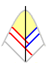
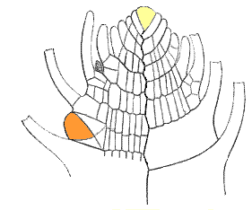
In this new row, the first diagram shows that the cells formed by the red and blue lines have grown in size, so the stem has increased in length. Moreover, there has been some increase in size of the apical cell as well. In the middle diagram you can see that a second red wall has formed, parallel to the first red wall. A third new cell has now been created. In the final diagram there is a second blue wall, parallel to the first blue wall, so that another new cell (the fourth) has been created. The creation of red and blue walls would continue in that same alternating sequence. In that final diagram the arrow indicates another development. In the first "red-wall" cell a cross-wall has formed and it divides the original "red-wall" cell into two cells. Of those two cells the one on the upper left is the outer cell and the other is the inner cell. The inner cell will remain a cell within the stem and will typically divide further. The outer cell may give rise to a side stem or a leaf. If, say, a side stem were to grow from that point the first thing to happen would be that a new apical cell would develop – the apical cell for the side stem. That apical cell would then cut off cells behind it in a manner similar to that already described and so the side stem would grow. Note that because the red and blue cells are cut out in sequence they are not symmetrical about the vertical axis through the midline of the stem. You can see this clearly in the next diagram, where the vertical axis is shown by the black line. Put simplistically, the bottom-most part of each blue cell is a little above the bottom-most part of the red cell that was cut out immediately before. The following diagram shows a longitudinal section of the apex area of a stem of the moss Fontinalis antipyretica. The diagram is Figure 49 from the third edition of JW Oliver's The Student's Introductory Handbook of Systematic Botany, published in London by Blackie & Son in 1898. By now quite a number of cells have been cut out from the apical cell and numerous additional cross-walls have formed in the inner cells to build up stem tissue. The curved, finger-like projections on either side of the stem are leaves. Towards the stem's apex the very immature leaves are shown in full. The leaves that are further away from the apex are more developed but in this drawing only the bases of those leaves are shown, the bulk of each leaf having been cut away in order to concentrate on the cells in the stem. The pale yellow cell indicates the apical cell of the main stem and the orange-brown cell indicates another apical cell from which a side-stem will start developing. The latter apical cell has developed from an outer cell in the main stem through the production of cross-walls, angled so as to produce appropriately oriented cutting faces. Apical cells are fundamental to gametophyte growth. Each stem, leaf, thallus branch and so on has its apical cell. Apical cells vary in shape and may have up to four cutting faces. Even the more detailed diagram of Fontinalis antipyretica shows a two dimensional view though stems are three dimensional. The apical cells in the stems of leafy bryophytes are often more-or-less tetrahedral, though often with somewhat curved edges. In the next pair of diagrams the one on the left shows such a tetrahedral apical cell, viewed at an oblique angle, in which three new cell walls have been created – red followed by blue and then black. The diagram on the right shows the new "red" cell pulled away a short distance to show you the newly created "blue" and "black" cells. As usual, the apical cell is in yellow.
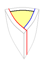
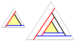
Apical cells and phyllotaxyThe following stylized diagrams show head-on views of a stem tip, with its tetrahedral apical cell. For simplicity, all edges are drawn straight rather than curved. These are snapshots taken at two points in time. On the left three cells have been cut out – first by the red wall, then by the blue and finally by the black. The view on the right shows the situation after the next three cells have been cut out, once again in the sequence red, blue, black. As in the earlier diagrams the coloured triangle shows the position of the apical cell after each trio of cuttings.
Look at the red-blue-black-red-blue-black sequence in the second diagram on the preceding line and imagine the process continuing. This process will lead to a row of red cells, a row of blue cells and a row of black cells along the length of the stem. Moreover the three rows would be symmetrically arranged around the stem, with each row 120 degrees away from the other two. In such a growth arrangement leaves on a stem would also be in three rows with the rows arranged symmetrically around the stem. It is very important to note two things. The first is that many leafy bryophytes don't show a 120° spacing between the rows of leaves. It is the case for the leafy liverworts but not for the great majority of mosses. There's more about mosses a little further down this page. The second thing to note is that though, in the above example, the rows would be symmetrically arranged around the stem the same cannot be said for the three leaves formed from any given red-blue-black trio of cells. Suppose you were able to take the row of leaves derived from the red cells and rotate the whole row 120° anticlockwise. Certainly that row would then be superimposed on the row of leaves obtained from the blue cells, but no red-row leaf would be superimposed on a blue-row leaf. Think back to the simple two-dimensional example given earlier. Those red and blue cells were cut out in sequence and were not symmetrical about the central vertical axis. Essentially, each blue cell was a little "higher" up the stem than the red cell that preceded it. The same holds true in the three-dimensional case. If, in the example given above, you wanted to superimpose each red-cell leaf on the blue-cell leaf that follows it, you'd need to rotate the red row 120° AND also move the whole row a little towards the stem's apex. Similarly when going from blue-cell leaves to black-cell leaves and from black-cell leaves to red-cell leaves and so on. The leaves are actually arranged in a spiral pattern but in such a way that, for any given leaf, the third successive leaf after it will be aligned with it and the three leaves have completed one circuit around the stem. Such an arrangement of leaves is said to have a phyllotaxy of 1/3 and that is common in the leafy liverworts. In the above diagrams the apical cells were drawn with the three sides of the front face of roughly equal lengths. That is by no means universal. In many of the leafy liverworts one side is markedly shorter than the other two, so that a much smaller cell is cut off along the corresponding cutting face. The majority of leafy liverwort species show a creeping, rather than upright, growth habit and the markedly smaller cutting faces are oriented towards the surface on which the liverwort is growing. Hence, if there are leaves of two different sizes growing from the stems, the smaller ones will be found on that side of the stem that faces the substrate. The leaves on that side of the stem are called the underleaves. In many moss species the stem's apical cell is more towards egg-like than tetrahedral, though still with three cutting faces, but with successive cells cut out at an angle that gives a phyllotaxy of 2/5. That is, the fifth leaf after a given leaf will be aligned with it and those five leaves complete two circuits around the stem. This also means that there are five rows of leaves along the stem. A number of genera have leaves in two rows, and hence a phyllotaxy of 1/2. Perhaps the most common of these is Fissidens, though in this genus the apical cell at a stem tip is initially tetrahedral, with three cutting faces, but it soon becomes lenticular, with just two cutting faces. There are also species with a 1/3 phyllotaxy, such as Fontinalis, which featured earlier. Some of the moss species with a 1/3 phyllotaxy are reminiscent of liverworts in that the leaves in one row are markedly smaller than the leaves in the other two rows. Other phyllotaxies amongst the mosses are 3/8, 5/13 and 8/21. Some other pointsIn the thallose bryophytes the apical cell may have two to four cutting faces. In a number of species the apical cell is wedge-shaped and cuts out cells alternately to the left and right, thereby increasing the size of the sheet-like thallus. At the other extreme cells are cut out left, right, top and bottom. Since apical cells are important it's no surprise to learn that they are protected. Gametophyte tissue in the area around the apical cell often grows more rapidly so that in leafy bryophytes there is a rapid growth of leaves around the apex which cover the apical cell. In thallose bryophytes the neighbouring areas of the thallus are often a little ahead of the apical cell, which is therefore contained within a slight protective notch. In addition there are often mucilage-producing cells near the apical cell which ensure that the apical cell does not dry out. An acrocarpous moss is one in which the female sex organs, or archegonia, are produced at stem tips. Stem tips that have produced female sex organs cannot continue growing since the apical cells in those stem tips have been used up in forming the archegonia. Hence there can be fresh growth from that stem only if a new apical cell develops below the old apex and generates a side-branch. Acrocarpous mosses generally show a tufty, upright growth habit and are sparingly branched. Pleurocarpous mosses are those in which archegonia are produced on short side branches. Pleurocarpous mosses typically grow in a creeping fashion, forming wefts or mats over the substrate, and are usually highly branched.
|