![An Australian Government Initiative [logo]](/images/austgovt_brown_90px.gif)

Life cycle – Sporophyte development
Hornworts
Hornwort sporophytes lack setae. Each tapering "horn-like" sporophyte, ![]() that grows out from a bulbous foot embedded in the thallus is entirely spore capsule. To see the foot you need to dissect the hornwort. The immature sporophyte is green and so, like the immature moss sporophyte, photosynthesizes.
that grows out from a bulbous foot embedded in the thallus is entirely spore capsule. To see the foot you need to dissect the hornwort. The immature sporophyte is green and so, like the immature moss sporophyte, photosynthesizes.
A liverwort or moss capsule comes to a definite stop in its development, with the spores having all matured together. The capsule then opens in some way to release the spores. In the hornwort genus Notothylas the sporophyte also has a determinate period of growth. By contrast in the other hornwort genera the sporophyte is continually growing from near the base. In theory such a sporophyte could grow indefinitely, though the death of the aging thallus or changes in environmental conditions eventually put a stop to sporophyte growth. Technically the near-basal area in which there is continual cell addition is an example of a meristem. Meristems are found in all plants for all plant growth is via meristematic tissue of some sort. The apical cell, mentioned in the introductory LIFE CYCLE page is an example of a meristematic cell.
The meristematic tissue near the base of a hornwort sporophyte is composed of a number of cells. By the production of new cells the meristem is constantly causing the sporophyte to extend upward. This means that the oldest part of the sporophyte is at the apex with regions progressively younger as you go down the sporophyte. When the sporophyte is still very short it is enclosed within a protective sheath (an involucre) but the sporophyte grows through it, leaving a cylindrical remnant around the sporophyte's base. This photo ![]() of a species of Megaceros shows some involucral remnants quite well.
of a species of Megaceros shows some involucral remnants quite well.
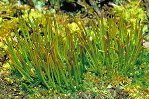 Phaeoceros, maturing sporophytes |
Given that the oldest cells in the sporophyte are near the apex it is in the apical region that the first spores mature. Then the spores a little lower mature and so on. In this photo the majority of sporophytes are brown in the upper region and green below. The spores in the brown areas are approaching maturity but in the green areas spore development is at various earlier stages.
As the spores mature the sporophyte walls change from green to brown, though mature spores may be green to brown, the colour depends on genus. When spores near the apex are mature the sporophyte develops slits there. Generally there are two slits but in some species there is only one and there are also species in which four slits develop. As the sporophyte dries the slits open and the spores can be released. As lower areas of the sporophyte mature the slits extend downward. This photo ![]() shows a number of sporophytes with dried and open upper regions. In many hornworts the slits stop a little before the sporophyte apex so that the sporophyte segments are united at their apices. In a mature sporophyte you will also find pseudo-elaters. These are tiny, filamentous structures that superficially resemble the spiralled elaters that are a feature of the capsules in many liverwort species. However, elaters and pseudo-elaters have different origins and there's more about the differences between elaters and pseudo-elaters in the ELATERS page. The pseudo-elaters are not spiralled in the genera Anthoceros, Folioceros, Phaeoceros, always spiralled in Megaceros and Dendroceros and have rudimentary spiral thickenings in Notothylas.
shows a number of sporophytes with dried and open upper regions. In many hornworts the slits stop a little before the sporophyte apex so that the sporophyte segments are united at their apices. In a mature sporophyte you will also find pseudo-elaters. These are tiny, filamentous structures that superficially resemble the spiralled elaters that are a feature of the capsules in many liverwort species. However, elaters and pseudo-elaters have different origins and there's more about the differences between elaters and pseudo-elaters in the ELATERS page. The pseudo-elaters are not spiralled in the genera Anthoceros, Folioceros, Phaeoceros, always spiralled in Megaceros and Dendroceros and have rudimentary spiral thickenings in Notothylas.
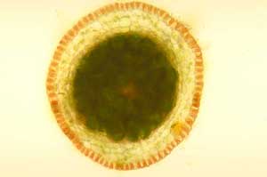 Cross section of Megaceros sporophyte |
This photo ![]() shows a cross section through the lower, still green region of a hornwort sporophyte. The yellow-green cells forming a ring around the centre are the sporogenous cells that will give rise to spores as well as the pseudo-elaters. Between the sporogenous cells and the sporophyte surface are the chlorophyllous, photosynthesizing cells. The photo (right) is of a cross-section of the upper part of a hornwort spore capsule that has started to turn brown. You can see that the outermost cells now have a brownish tinge. Where the previous photo showed sporogenous cells there is now a mass of dark green cells – a mix of mature spores and pseudo-elaters. Here
shows a cross section through the lower, still green region of a hornwort sporophyte. The yellow-green cells forming a ring around the centre are the sporogenous cells that will give rise to spores as well as the pseudo-elaters. Between the sporogenous cells and the sporophyte surface are the chlorophyllous, photosynthesizing cells. The photo (right) is of a cross-section of the upper part of a hornwort spore capsule that has started to turn brown. You can see that the outermost cells now have a brownish tinge. Where the previous photo showed sporogenous cells there is now a mass of dark green cells – a mix of mature spores and pseudo-elaters. Here ![]() are some spores and pseudo-elaters.
are some spores and pseudo-elaters.
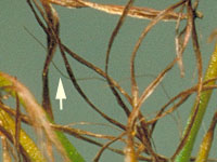 Along the hornwort sporophyte's central axis there is a column of sterile tissue called a columella. This is thought to be a conducting system that helps supply nutrients to the developing spores. When you look at a group of open sporophytes it's easy to see the capsule segments but amongst them you'll find the columellas, thinner and wiry in appearance. If you use a handlens to look closely at mature sporophytes such as the ones shown in this earlier photograph
 ![]() you will be able to see columellas amongst the split capsules. The columellas are markedly thinner than the sporophyte segments. On the left is an enlargement of part of that previous photo and the arrow points to a columella.
you will be able to see columellas amongst the split capsules. The columellas are markedly thinner than the sporophyte segments. On the left is an enlargement of part of that previous photo and the arrow points to a columella.