![An Australian Government Initiative [logo]](/images/austgovt_brown_90px.gif)

Two Groups - classifying fungi into ascomycetes and basidiomycetes:
Gasteromycetes
In all of the other basidiomycetes the basidia are persistent. Even by the time a mushroom has largely rotted away you can usually still find basidia on the gills. There are several common groups of basidiomycetes where the mature fruiting body has spores - but no basidia. To find the basidia you must have an immature (but not too young!) fruiting body, because once the spores have been produced the basidia break down. Examples of such fungi are various powdery spored fruiting bodies (the puffballs and similar fungi), the slimy-spored stinkhorns and the Bird's Nest Fungi.
These are examples of a subgroup of the basidiomycetes commonly called the gasteromycetes. The word gasteromycete literally means "stomach fungus" - and these fungi produce their spores inside the fruiting body that, at least initially, is enclosed within an outer skin. While, as a group, these fungi may all be referred to as gasteromycetes, that name does not signify close evolutionary relationships between all the members. The name is simply one of convenience - rather than being one of classificatory importance. Reflecting their diverse origins, these fungi show considerable variation in both the overall appearance and internal structure of the immature, basidia-bearing fruiting bodies. Many start as more-or-less spherical masses of homogenous tissue, and then develop various patterns of internal spore-bearing areas. In some species these spore-bearing areas consist of empty cavities, with the basidia lining the walls and protruding into the empty interiors. In others the spore bearing areas may be filled with basidia and hyphae or a gelatinous matrix while the spores are developing.
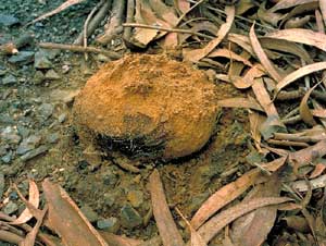
Pisolithus is a common powdery-spored gasteromycete genus, with the
fruiting bodies often seen pushing up through bitumen roads ![]() .
.
 |
cross-section (right) |
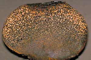 |

A vertical cross-section of such a fruiting body shows a powdery region at the top, but with the lower part looking as though it consisted of a multitude of densely packed rice grains (called peridioles), comprising packets of spores. The peridioles are relatively thin skinned and the whole mass is contained in a brittle outer skin. This highly stylized diagram shows a vertical cross-section through a very immature Pisolithus fruiting body. The black circle represents the outer skin, the internal tissue is in brown and there are numerous chambers (shown in white). Each of these chambers eventually becomes the interior of a peridiole. These chambers are isolated from each other and contain the basidia. The chambers fill quickly with a gelatinous matrix, the spores become detached from their basidia and complete their development (after detachment) within the gelatinous interior. Of course, in most cases people only see the Pisolithus fruiting bodies in the advanced, powdery stage as shown in the photograph.
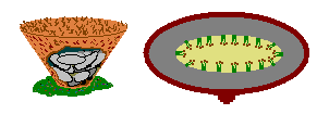 Cyathus novae-zealandiae (right) |
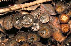 |
The mature fruiting body of a Bird's Nest Fungus (such as this Cyathus novae-zealandiae) consists of a leathery, cup-like structure that contains a number of disc-shaped "eggs" (peridioles again). These peridioles differ from the Pisolithus peridioles by being enclosed in a thick outer casing and by becoming separated from each other. The cup-like structure is usually no more than a centimetre in diameter at the top. Before the Cyathus fruiting bodies are mature, they have a membrane across the top of the cup. You can see a couple of immature fruiting bodies at the right in the photograph. The diagram on the left shows an immature cup, cut away to show it full of peridioles. The middle figure shows the basidia in the central part of one such peridiole. The thick brown line represents the peridiole's hard outer casing. At a young stage, the interior of a peridiole has a gelatinous filling and at an even younger stage the fruiting body would have had the same type of chambered appearance as shown in the stylized diagram for Pisolithus.
The preceding cross-section diagram shows a very simple type of internal structure, but numerous gasteromycetes have much more complicated patterns of development.
 Many
mature puffballs have a very simple structure. There's just a thin-walled sack
of powdery spores with a hole at the top, through which the spores are puffed
out when the sack is compressed. This simplicity at maturity belies the complex
internal structure of the immature fruiting body during spore production. At
that stage the fruiting body has a very convoluted interior. The diagram represents
a stylized vertical cross-section of such a young puffball. Here the cavities
are not formed as separate chambers, but within ingrowths of tissue from the
periphery. The basidia develop on those convoluted surfaces, shown here in brown.
Many
mature puffballs have a very simple structure. There's just a thin-walled sack
of powdery spores with a hole at the top, through which the spores are puffed
out when the sack is compressed. This simplicity at maturity belies the complex
internal structure of the immature fruiting body during spore production. At
that stage the fruiting body has a very convoluted interior. The diagram represents
a stylized vertical cross-section of such a young puffball. Here the cavities
are not formed as separate chambers, but within ingrowths of tissue from the
periphery. The basidia develop on those convoluted surfaces, shown here in brown.
 See colour coding in diagram to lower left of page. |
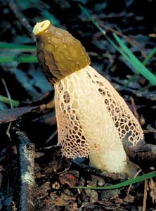 Dictyophora multicolor |
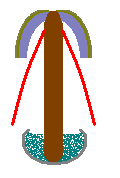
The fruiting bodies of stinkhorns in the genera Dictyophora, Mutinus
and Phallus consist of a stem with the slimy spore mass on a small cap
at the top of the stem. The cap isn't spread out (like a typical mushroom) but
hugs the stem fairly closely. In this picture of Dictyophora multicolor
you can see the small, slimy cap. In addition, there is a mesh-like skirt that
hangs down below the cap. This picture of a dissected Phallus rubicundus
![]() shows the cap a little more clearly.
shows the cap a little more clearly.
 In
line with the style of the two gasteromycete diagrams above, the diagram on
the left represents a vertical cross-section of a young stinkhorn from any of
the three genera listed above. You can see the unexpanded stem, the curved cap
and the short projections (growing in from the marginal tissue) that bear the
basidia.
In
line with the style of the two gasteromycete diagrams above, the diagram on
the left represents a vertical cross-section of a young stinkhorn from any of
the three genera listed above. You can see the unexpanded stem, the curved cap
and the short projections (growing in from the marginal tissue) that bear the
basidia.
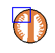
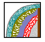 It
is important to realize that these diagrams are very simplistic, two-dimensional
representations of considerable three-dimensional complexity. Looking more closely
at the young stage of Dictyophora, here is a more detailed (but still
stylized) diagram of the structure, showing an enlarged view of the area contained
within the blue rectangle above. The young stinkhorn is enclosed within a thin,
leathery skin, represented here by the thin grey line. Immediately beneath that
skin is a relatively broad gelatinous zone (the stippled band). Next are the
finger-like to plate-like projections (khaki green) that bear the basidia; the
body of the eventual cap (bluish); the tissue that will form the skirt (shown
here as a red net); other hyphal/gelatinous tissue (stippled pale grey) and
the stem (the brown vertical bar at the right). The projections can form quite
intricate channels and chambers but (once the spores have matured) they break
down into the smelly, khaki to brown slime that holds the spores on the outside
of the cap of the mature fruiting body.
It
is important to realize that these diagrams are very simplistic, two-dimensional
representations of considerable three-dimensional complexity. Looking more closely
at the young stage of Dictyophora, here is a more detailed (but still
stylized) diagram of the structure, showing an enlarged view of the area contained
within the blue rectangle above. The young stinkhorn is enclosed within a thin,
leathery skin, represented here by the thin grey line. Immediately beneath that
skin is a relatively broad gelatinous zone (the stippled band). Next are the
finger-like to plate-like projections (khaki green) that bear the basidia; the
body of the eventual cap (bluish); the tissue that will form the skirt (shown
here as a red net); other hyphal/gelatinous tissue (stippled pale grey) and
the stem (the brown vertical bar at the right). The projections can form quite
intricate channels and chambers but (once the spores have matured) they break
down into the smelly, khaki to brown slime that holds the spores on the outside
of the cap of the mature fruiting body.
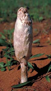 |
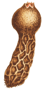
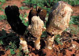 Podaxis pistillaris |
The stem-and-cap type of development (as shown above in the simple brown diagram) is not restricted to stinkhorns. The powdery spored Podaxis pistillaris also has such a form when young. In the mature stage Podaxis fruiting bodies look like powdery drumsticks. The powdery region is initially contained within a brittle, white outer skin, but the skin breaks away to expose the dark brown to black spore powder.
There are a few more forms of immature gasteromycete fruiting bodies than shown here, but the types shown here will give you some idea of the variety. As you can see, the gasteromycete fruiting bodies can be quite complex and to understand the complexity you need to follow the development of the fruiting body and, in particular, note where the basidia form.