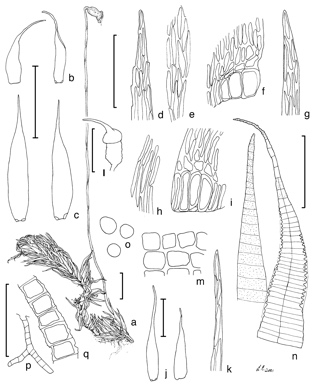
Caption: A, gametophyte and sporophyte. B, branch leaves of T. wattsii isotype. C, branch leaves of Hypnum glaucoviride isotype. D, leaf apical cells of B. E, mid-laminal cells of B. F, alar region of B. G, apical leaf cells of C. H, mid-laminal cells of C. I, alar region of C. J, perichaetial leaves. K, apex of perichaetial leaf. L, capsule with operculum. M, exothecial cells. M, peristome: endostome segment (left), exostome tooth (right). O, spores. P, branched gemma arising from the stem. Q, close-up of gemma (L, P, Q, I.G.Stone 21424 MEL). Scales: 1 mm for habit, leaves, capsules and gemmae; 50 ?m for spores; 100 mm for cellular drawings including peristome.
Reproduced from H.P.Ramsay, W.B.Schofield & B.C.Tan, The Journal of the Hattori Botanical Laboratory 95: 51, fig. 22 (2004). Illustrators: L. Eklan & H.P. Ramsay
Australian Mosses Online